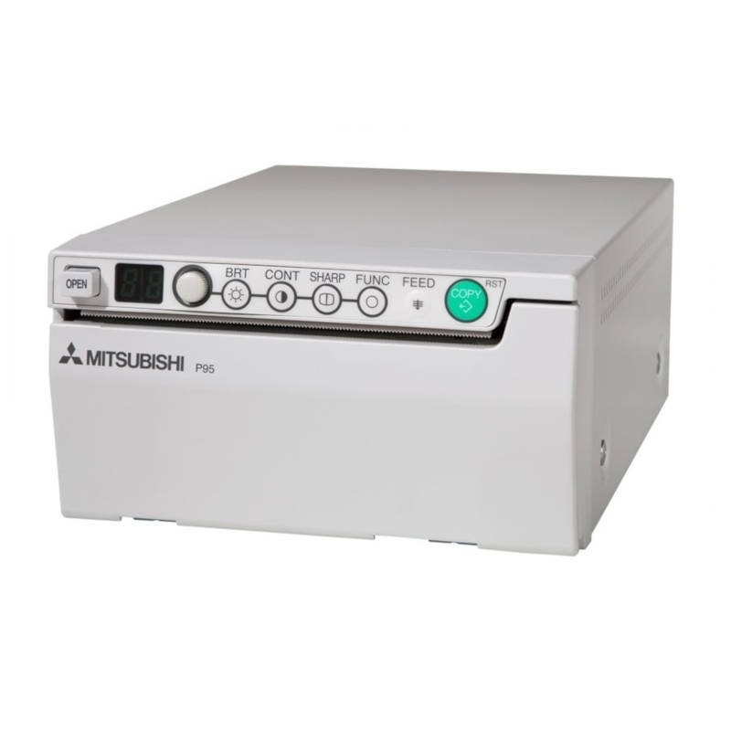
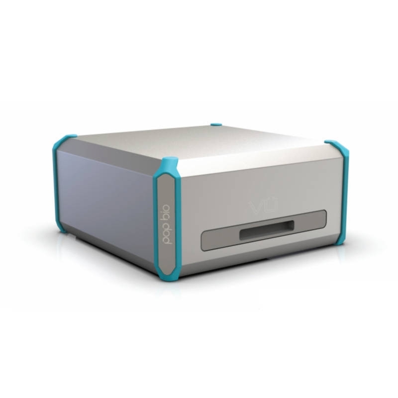
Vü-C Chemiluminescence System

VÜC

Clever engineers have taken the very latest imaging sensors and have adapted them for blot imaging. All this new technology is packaged into a compact device giving you a smart new way to capture your blot images.

Features :

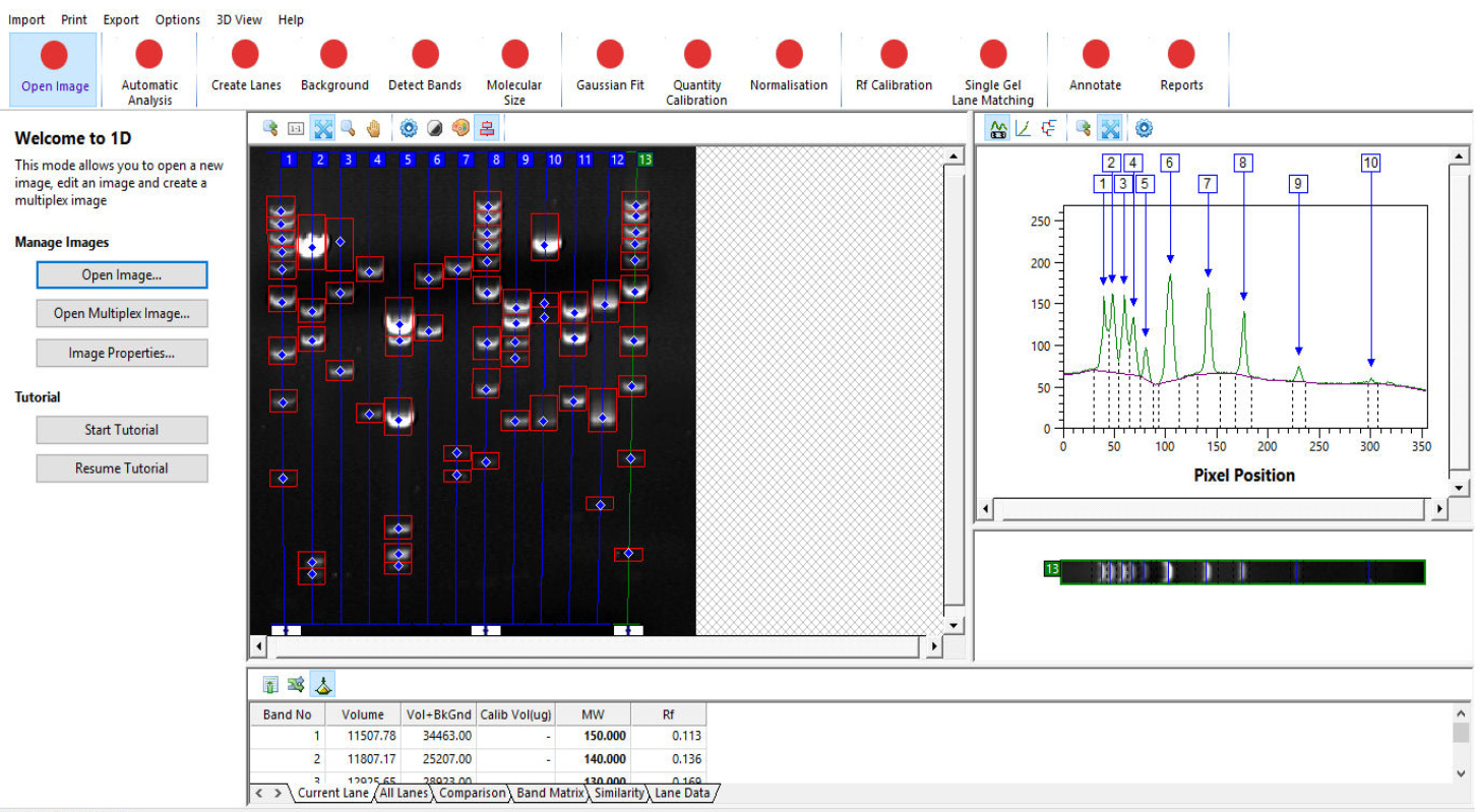
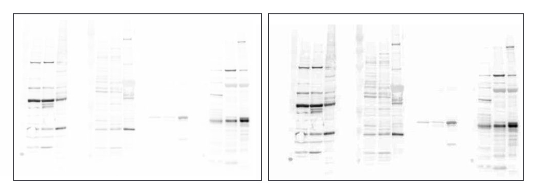
  • Western blot imaging with a difference
  • Ultra resolution
  • Super sensitivity
  • Stunning images
  • No camera, no lens, no setup

More details

Description
Specifications
Related products
Resources

Description

Using Advanced Progressive Imaging technology, the high sensitivity sensors can easily capture low output blots to give you an outstanding image. All you have to do is to select the tray size for your blot, place the blot in the marked position and push the tray into Vü with your finger. Vü immediately recognises the tray and then adapts to the blot size or application type. You will get your image in a matter of a few minutes.

Vü is a stunning new system which utilises a new imaging platform. It sits discreetly on any laboratory bench. Just use your cell phone, tablet, laptop or computer to collect the image and that's all you have to do. Depending on your sample type, your image will appear in seconds or minutes ready for you to print or analyse. No fuss, no hassle, no drama, no camera, lens or filters to worry about. No sensitivity issues, no focussing needed. Just the best image you have ever seen whether it is of a faint band or a high signal output blot.

Analysis software

Analysis of Western blots with Vü's software is rapid, automated to a high level and reproducible. Highly developed algorithms accurately detect lanes and bands even on distorted gel images. Calibrate the bands using one or more Molecular Size standard lanes and derive absolute band quantitation using known quantity calibration standards in your samples.

Comparative Images

Vü is as good, if not better, than a traditional CCD camera based gel and blot imaging system. See below and decide which image you prefer. The one on the left was captured using  a well known competitive system while the image in the right was acquired using a Vü-C. Both took the same amount of capture time. The competitive system costs 3 times that of the Vü-C and of course requires a lot more set up and control.

Which imaging device would like to see in your laboratory...?

Specifications

IlluminationRGB integral LEDS for marker including coloured markers
Sample size10 x 10 cm
Sensor coolingNot required
Sensor resolution51M pixels
Sensor lens T-Stop1.01
Dimensions (W x D x H)35.3 x 37.5 x 17.5 cm
Weight8 kg
Power supply100- 250V external power supply
ConnectivityIntegral Wifi and ethernet

Resources