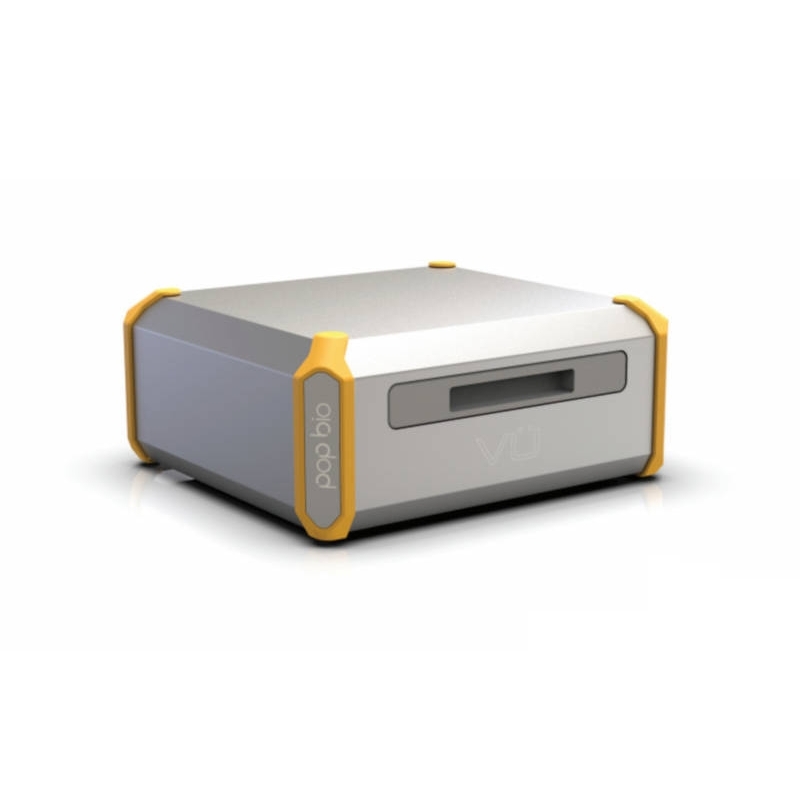
Vü-F Fluorescence System

VÜF

Isn’t it time to upgrade and use the latest technology to enhance your gel and blot imaging ? That’s why the Vü has been developed with the Advanced Progressive Imaging technology.

Features :

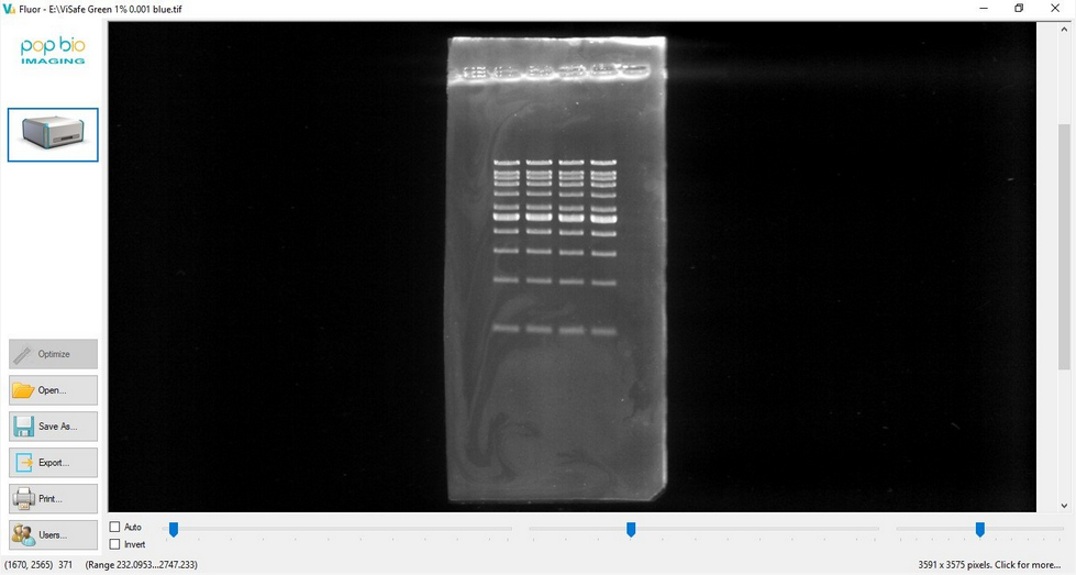
  • Fluorescence imaging made easy
  • One step operation
  • UV, blue and white light illumination
  • Stunning images
  • No camera, no lens, no filters, no lasers, no setup

More details

Description
Specifications
Related products
Resources

Description

Vü-F is for fluorescence applications such as DNA gels, protein gels, safe dye gels. The sliding drawer can accept samples up to 20 cm x 20 cm and has a series of trays available for different application types. Just place your sample on the tray and push it in. Vü-F detects the sample type and captures an image using either UV or blue light. The use of traditional ethidium bromide stained UV gels may be declining but there's still a 302 nm light source built in. For safe dyes there are integral blue LED’s and an option to have Blue/Green illumination. One can still use a traditional white light converter to view commassie and silver stain samples.

There is no fancy screen or massive processor into Vü-F which needs constant upgrading and can go wrong. Why build all that expense into a system when you probably already have better computing devices in your laboratory that can be utilized instead ? Maybe you just want to use a specific device or something your IT department is happy with. Either way with Vü-F you are in control.

With Vü-F you can take any mobile device, tablet, laptop, computer and link it with it. In fact, your only control is to capture the image – no fussy setups are required because there is no camera, lens or filter wheel. Just push the sample draw in and Vü-F does the rest.

Analysis software

Analysis of 1D gels with Vü's software is rapid, automated to a high level and reproducible. Highly developed algorithms accurately detect lanes and bands even on distorted gel images. Calibrate the bands using one or more Molecular Size standard lanes and derive absolute band quantitation using known quantity calibration standards in your samples.

Specifications

UV illuminationIntegral 302 nm
Blue light source
Integral blue LED's
Blue/green lightOptional
Sample size20 x 20 cm
Sensor resolution90M pixels
Sensor lens T-Stop2.86
Dimensions (W x D x H)42.6 x 48.0 x 20.5 cm
Weight15 kg
Power supply100- 250V external power supply
ConnectivityIntegral Wifi and ethernet

Resources